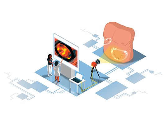
The research group is working on the development of minimal and non-invasive imaging-based assistance systems for surgical applications. The aim is to provide the surgeon with anatomical or physiological information that is not visually detectable, such as the representation of tissue in the depth and the visualization of temperature changes or tissue perfusion. The group focuses on the following imaging techniques: ultrasound imaging to support brain tumor surgery, infrared thermography for skin graft planning, hyperspectral imaging to monitor patient condition, identify anatomical structures and assess tissue perfusion in visceral surgery and optical coherence tomography in dentistry and oncology. Besides the clinical evaluation and improvement of the intraoperative imaging devices, innovative and specific algorithms are developed to support the interpretation and analysis of the image data. The group works in close collaboration with clinical partners of the Leipzig University Hospital and industrial partners.
Research goals
1) The clinical and objective evaluation of new imaging modalities in the operating room for specific applications. For this, image data are acquired during surgical interventions of patients and evaluation methods are developed. This first goal is carried out in close cooperation with our partners of the Leipzig University Hospital.
2) The further development of imaging devices for optimal use in the operating room under operating conditions (limited space, sterile environment). For example, the development of systems for capturing 3D data and the miniaturizing of devices are current issues. Industrial partners are involved in such projects.
3) The development of computer-assisted tools to support the interpretation and analysis of image data. The image and signal data do not contain intuitive information (temperature, light spectra) and are difficult to interpret. Therefore, a further objective is the development of registration, segmentation and visualization methods for the automatic detection, enhancement and classification of tissue and anatomical structures.
Research topics
Former and current projects include the following topics:
The automatic model-based segmentation of blood vessels and brain tumor tissue in intraoperative 3D ultrasound image data in neurosurgery;
The automatic detection of skin perforators in static and dynamic thermography in plastic surgery;
The evaluation of skin perfusion in infrared thermography during reconstruction surgery;
The detection and evaluation of carious lesions using optical coherence tomography;
The tissue classification and the evaluation of organ perfusion using hyperspectral imaging in visceral and thoracic surgery.
Teaching
The scientists of the group are involved in courses for master students of computer science (Leipzig University and HTWK) and students of human medicine (Leipzig University).