


Prof. Dr. habil. Claire Chalopin

The aim is to develop a new laparoscopic imaging system and supporting non-invasive intraoperative measures to increase the identification and classification of risk structures and lesions for visceral, transplantation, thoracic and vascular surgery based on the latest high-resolution HSI technology.
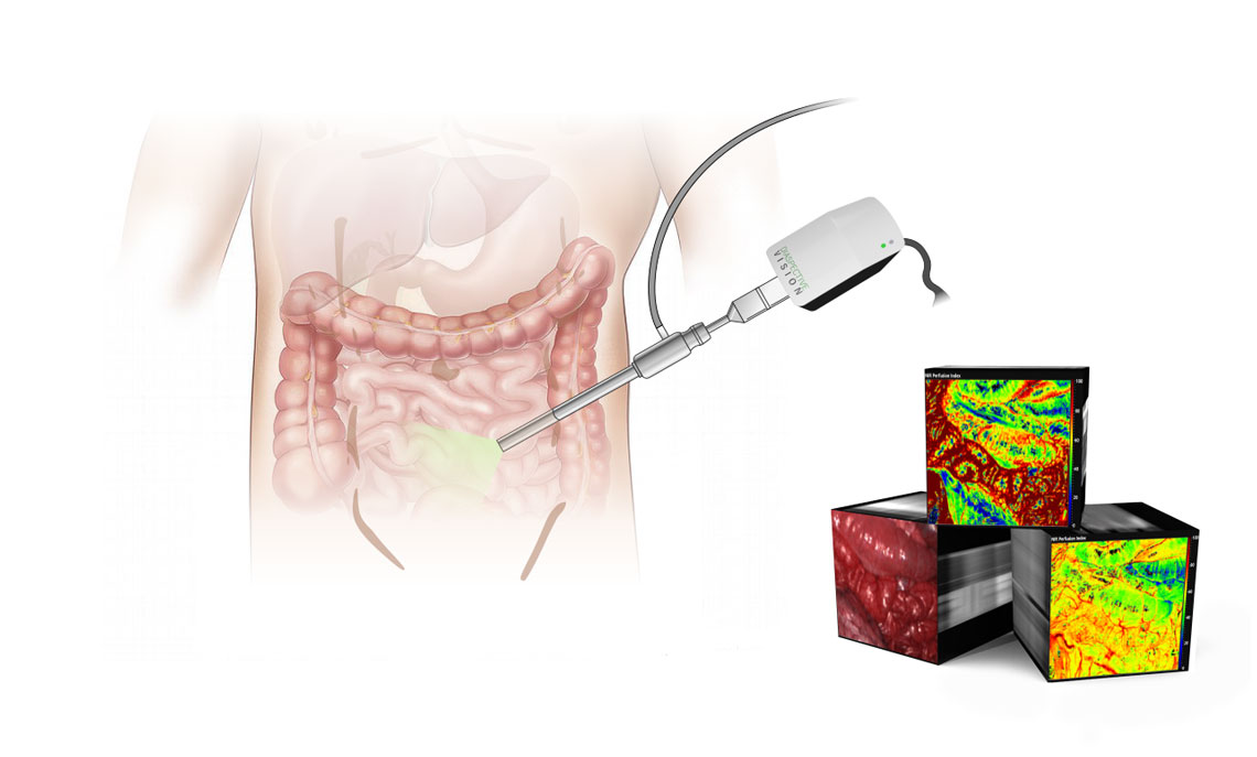
Laparoscopic surgery plays an important role in the surgical oncology and visceral surgery. However, the field of view of the surgeon through the laparoscope is limited and the accurate identification of the anatomical risk structures and lesions depends on the surgeon’s experience. Therefore, additional imaging modalities were added to support the surgeon in this complex task. For example, ultrasound imaging is standardly used for the localization and examination of lesions. However, this technology is not suitable for hollow organs such as the bowel. Further methods are the tattooing of tumors and the fluorescence imaging. The limitations are the use of contrast agents and the inaccurate delineation of the tumor margins.
Hyperspectral imaging (HSI) is a relatively new imaging method in medicine. The principle combines imaging with spectrometry. This modality showed promising results for the identification of structures and the assessment of tissue perfusion in the research area in the visceral and thorax surgery.
In this context, the purpose of the LYSiS project is the development of a new intraoperative laparoscopic imaging system to support the identification and classification of risk structures and lesions in the visceral and thorax surgery based on the non-invasive HSI technology.
The developed system will be evaluated on ex-vivo tissue samples resected during patient operations. Based on the first results, an optimization of the system for intraoperative applications directly on patients and towards the commercialization of the future product will be done.