


03.07.2019
 Newsletter on 2nd quarter
The second ICCAS Newsletter 2019 reports on latest trade fair and congress activities, presents current publications and invites to upcoming events such as the next ICCAS Open Day. (read more)
28.06.2019
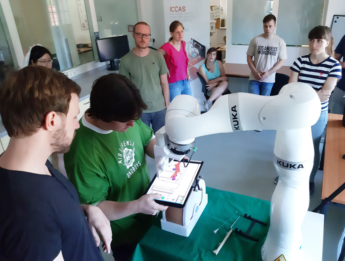
Informatic students test ICCAS-developments
Medical informatics students from universities in Brunswick, Heidelberg and Amsterdam received an insight into the research in computer-assisted and robot-aided surgery (read more)
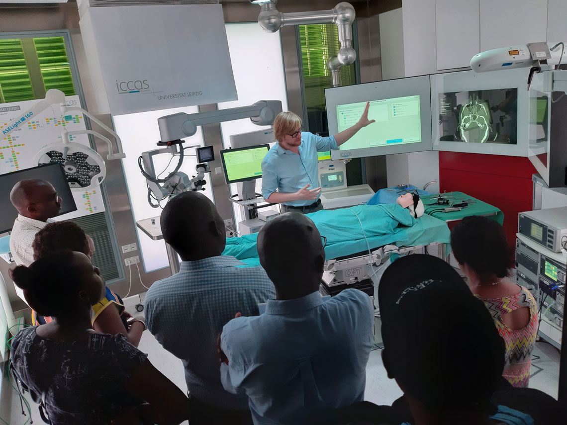
Kenyan partner universities at ICCAS
On June 26, teachers from two Kenyan partner universities visited us to learn more about practice-oriented university teaching and application-targeted project work. (read more)
27.06.2019
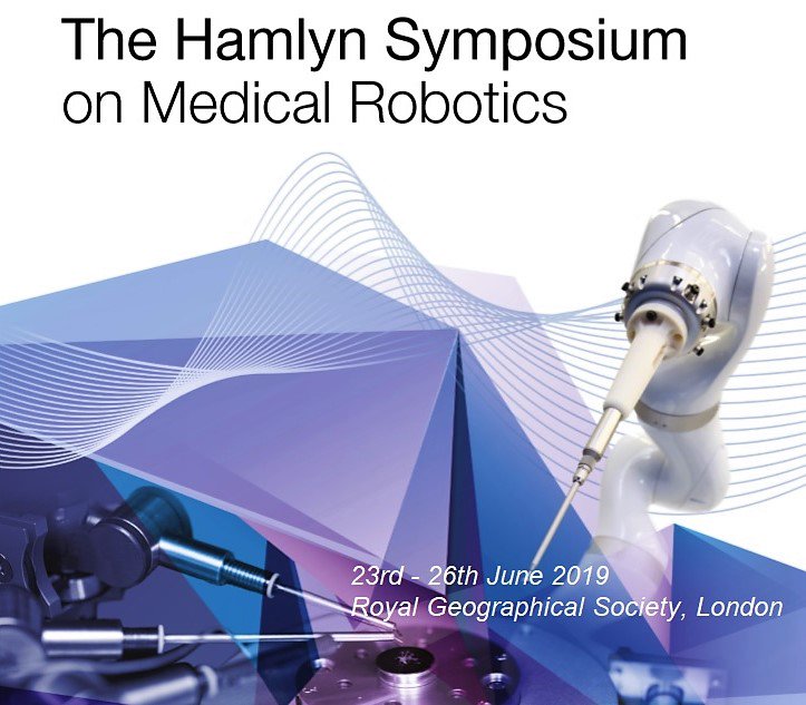
Presentation of new MR-guided procedures
Prof. Andreas Melzer gave a lecture on new technologies for MR-guided endovascular procedures at the renowned Hamlyn Symposium on Medical Robotics (HSMR) (read more)
20.06.2019
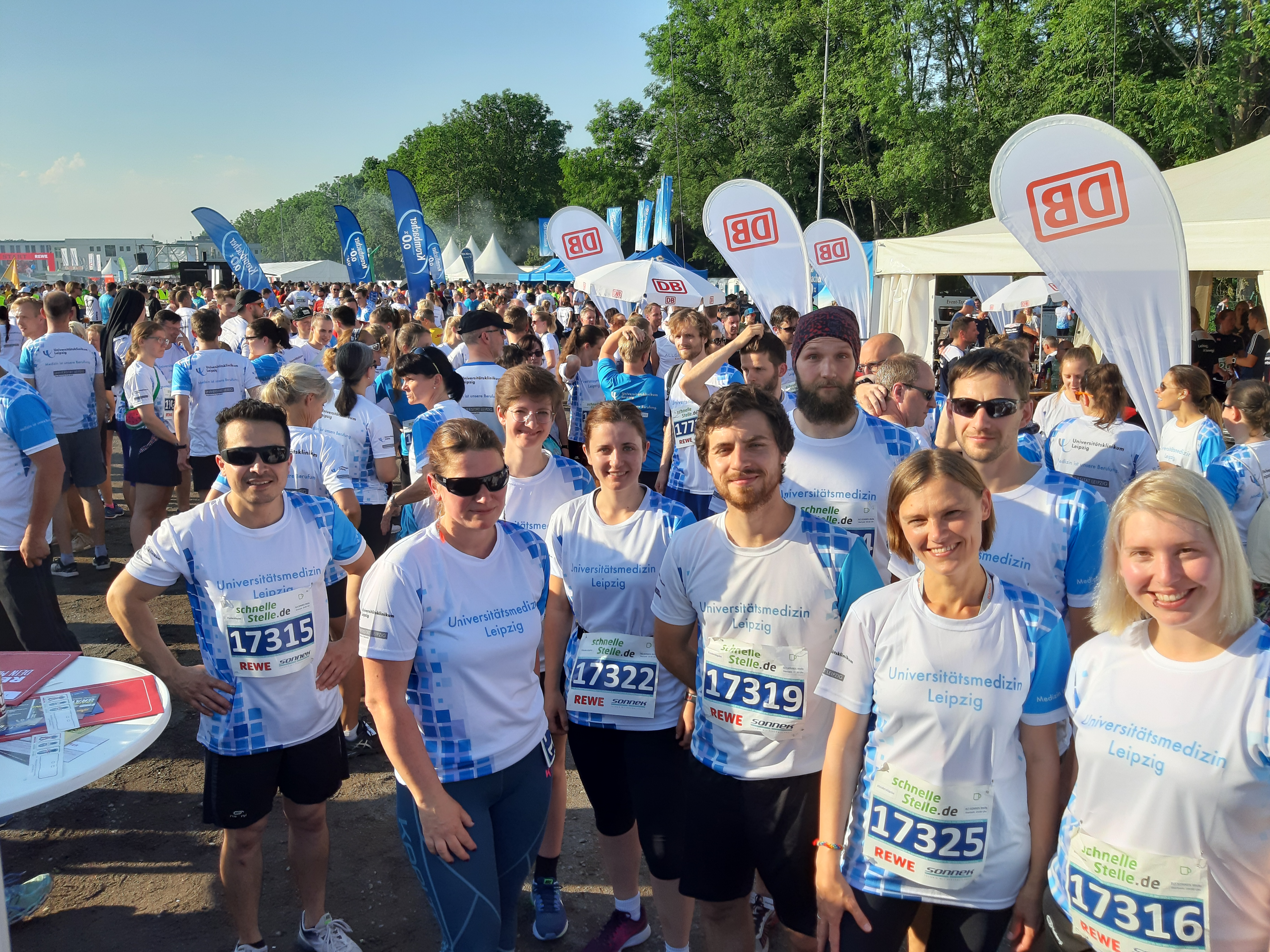
ICCAS shows its sporty side
ICCAS once again took part in the Leipzig Corporate Run for University Hospital and Faculty of Medicine yesterday. Under tropical conditions, the ICCAS team focused not only on personal best times but also on team spirit and sporting fun. With over 20,000 runners from 1,000 companies, this year’s event achieved a new participation record. Thanks to more than 600 running enthusiasts, the University Medicine was once again awarded the title “Most Sporty Company”.
19.06.2019
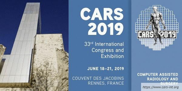
ICCAS presents results at CARS Congress
At this year’s Computer Assisted Radiology and Surgery – Congress (CARS), which takes place in Rennes (France) from June 18 to 21, the MAI team enriches the IFCARS / SPIE / ISCAS Joint Workshop “Digital Operating Room”: Prof. Thomas Neumuth gives an invited lecture on artificial intelligence and machine learning in the operating room of the future. Johann Berger and Juliane Neumann present research results on the networking of medical devices based on IEEE and the SDC interface.
Marianne Maktabi (MIB group) talks about tissue classification based on hyperspectral data during a further congress session.
13.06.2019
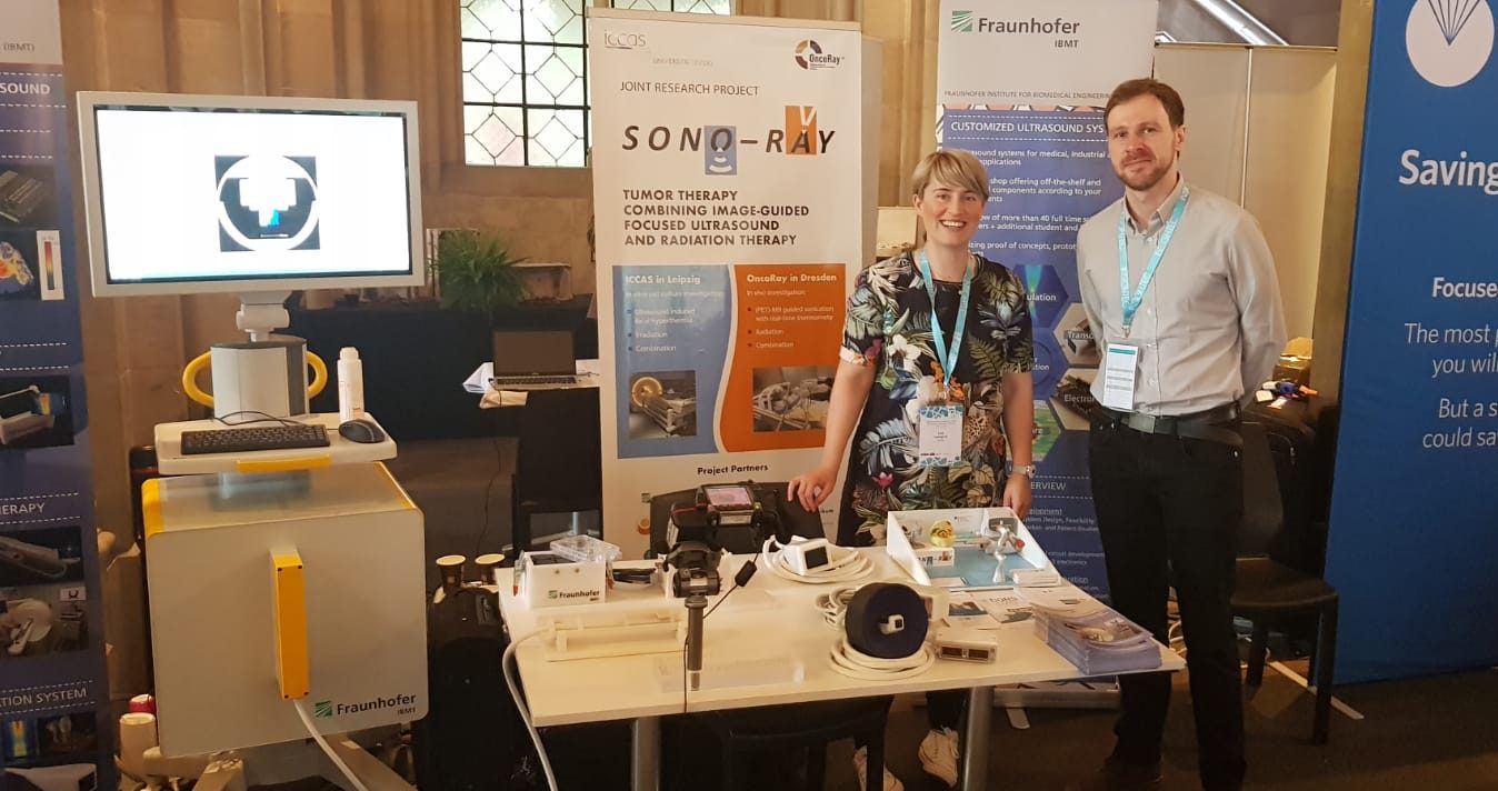
SONO-RAY group exhibits at ISTU / EUFUS Joint Symposium
At the Joint International Symposium for Focused and Therapeutic Ultrasound (EUFUS/ISTU) in Barcelona (Spain), scientists from the SONO-RAY project present results on robot-assisted and MR-guided focused ultrasound hyperthermia at the exhibition booth and in poster presentations. Furthermore, project leader Prof. Andreas Melzer acts as congress co-chair and chair of two panels. Group leader Dr. Lisa Landgraf is chair of the session ‘Therapy Ultrasound’.
12.06.2019
Robotics theme and SDC presented at EAES Congress
This year’s 27th Congress of the European Association for Endoscopic Surgery (EAES) takes place in Sevilla (Spain). Johann Berger will present the results of the work on “Modelling a Collaborative Robot with the IEEE 11073 SDC Standard for Combined Focused Ultrasound and Radiation Therapy” at the Technology Symposium, on June 12. On June 14, Martin Kasparick and Prof. Thomas Neumuth from the OR.NET e.V. present the OR.NET-EAES declaration on medical device interoparability to surgeons.
28.05.2019
SONO-RAY-results presented at German Congress of Radiology
The “100. German Congress of Radiology” is the next specialist forum where the results of the SONO-RAY-group will be presented. Here, Dr. Lisa Landgraf is holding a lecture on the successful radiosensitization of tumor cells in vitro by Focused Ultrasound Hyperthermia (FUS-HT) on May 31.
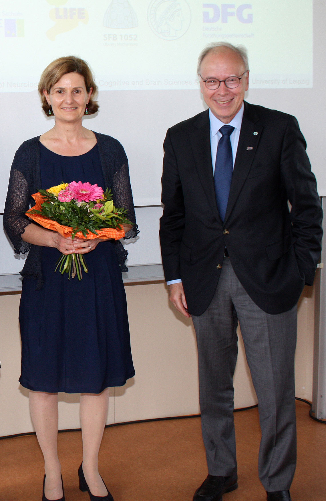
24.05.2019
Successful habilitation for Dr. Claire Chalopin
Dr. Claire Chalopin successfully completed her habiliation process. Congratulations!
Her postdoctoral thesis deals with the technical development of intraoperative 3D ultrasound with contrast medium and dynamic infrared thermography (DIRT) for neurosurgery. The imaging technique shows promising results for the visualization of structures and the assessment of tissue perfusion, but needs to be more optimally integrated into the surgical workflow. In addition, computer assistants will support the analysis of the imaging data.